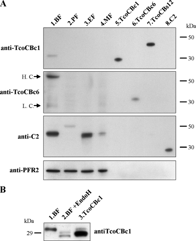
FIG. 5.
Western blot analysis of TcoCB expression in T. congolense. (A) Proteins from 2 × 10⁶ bloodstream, procyclic, epimastigote, and metacyclic forms (lanes 1 to 4, respectively) were separated on SDS-PAGE under reducing conditions together with 10 ng of recombinant proteases TcoCBc1, TcoCBc6, pro-TcoCBs12, and C2 (lanes 5 to 8, respectively), transferred to a PVDF membrane, and probed sequentially with antisera raised against different cysteine proteases. PFR2 was used as a loading control. Arrows indicate mouse IgG heavy chains (H.C.) and light chains (L.C.) recognized by the anti-mouse IgG peroxidase-conjugated secondary antibody. (B) Deglycosylation analysis of T. congolense bloodstream forms. A total of 2 × 10⁶ bloodstream forms were left untreated (lane 1) or were treated (lane 2) with endoglycosidase H, and proteins were separated by SDS-PAGE together with 10 ng of recombinant nonglycosylated TcoCBc1 (lane 3). Proteins were transferred to a PVDF membrane and probed with antiserum raised against TcoCBc1.

