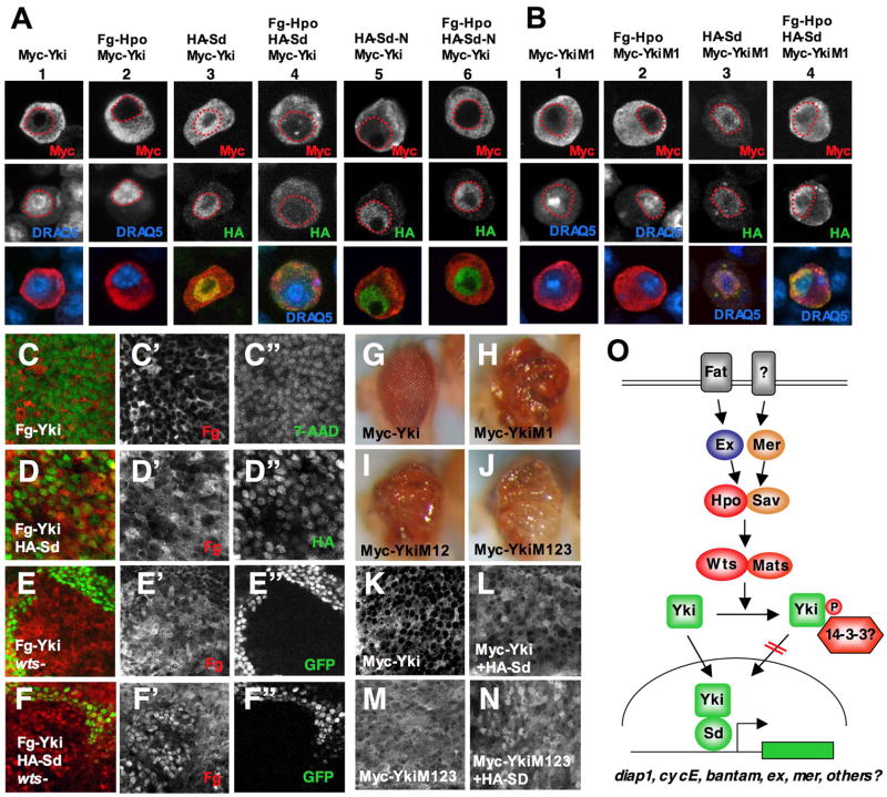
Figure 7. Hpo signaling regulates Yki nuclear localization and activity through phosphorylating S168.
(A–B) S2 cells expressing Myc-Yki (A) or Myc-YkiM1 (B) with or without Fg-Hpo and/or HA-Sd were immunostained with anti-Myc (red) and anti-HA (green) antibodies. Nuclei were marked by DRAQ5 (blue) and demarcated by red dashed lines. Images in individual channels were shown as black and white in top and middle rows and merged images in bottom rows. (C–D″) High magnification view of wing discs expressing Fg-Yki either alone (C–C″) or together with HA-Sd (D–D″) using MS1096 and immunostained with anti-Flag (red) and anti-HA (green in D) antibodies. The nuclei were labeled by 7-AAD (green in C–C″). (E–F″) High magnification view of wing discs carrying wts clones and expressing Fg-Yki either alone (E–E″) or together with HA-Sd (F–F″). wts mutant cells were marked by the lack of GFP expression (green). (G–J) Adult eyes expressing Myc-Yki (G), Myc-YkiM1 (H), Myc-YkiM12 (I), or Myc-YkiM123 (J) with GMR-Gal4. Of note, a strong transgenic line for Myc-Yki is shown here. (K–N) High magnification view of wing discs expressing Myc-Yki (K), Myc-Yki plus HA-Sd (L), Myc-YkiM123 (M), or Myc-YkiM123 plus HA-Sd (N). (O) The Drosophila Hpo pathway. See text for details.

