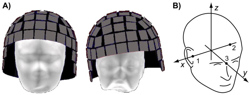
Figure 1.
Head position within MEG sensor array. A) The scalp surface for an adult (left) and a child (right) subject is shown within a representation of a helmet-shaped array of MEG sensor elements (gray squares). The sensor array is immersed in liquid helium Dewar (not shown), which imposes a minimum distance between the head and the sensor array. However, there is typically room for the head to move with respect to the sensors, especially for children with a small head. B) The head-based coordinate system, defined by three anatomical landmarks: right and left pre-auricular points (1 & 2) and nasion (3).

