Figure 1.
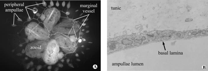
(A), Colony of Botryllus schlosseri seen in vivo from the ventral side. The vasculature has been injected with fluoresceine in order to highlight the peripheral vessels and the ampullae. (B), Electron micrograph showing the baso-apical structure of a regenerated peripheral vessel after surgery. Magnification 14,000X.

