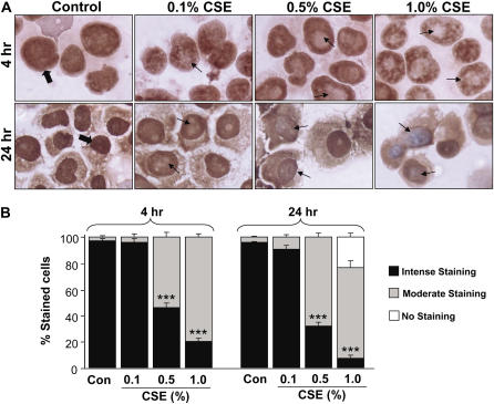
Figure 5.
Decreased sirtuin (SIRT1) protein staining in response to cigarette smoke extract (CSE) treatment in monocyte–macrophage (MonoMac6) cells. (A) CSE decreased the levels of SIRT1 in MonoMac6 cells at 4 and 24 hours. MonoMac6 cells were treated with different concentrations of CSE (0.1–1.0%). Cells were harvested and the cytospin slides were prepared at 4 and 24 hours of treatments. Immunostaining was performed using a rabbit polyclonal antibody specific for SIRT1 followed by the avidin–biotin–peroxidase complex method, and counterstained with hematoxylin. The dark brown color represents the presence of SIRT1, which was decreased in response to CSE treatment. (B) Graph showing the percentage of SIRT1-positive cells from the total number of cells in CSE-treated MonoMac6 cells. The assessment of immunostaining intensity was performed semiquantitatively and in a blinded fashion. Results shown are means ± SEM of three separate experiments (n = 3). ***P < 0.001, significant compared with control values.

