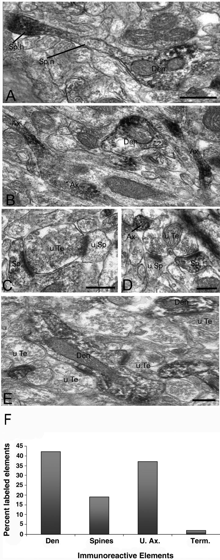
Figure 8.
Ultrastrutctural analysis of R7BP-immunoreactive neuronal elements in the rat striatum (A-D) and ventrolateral thalamus (E). A) Immunoreactive dendritic shaft (Den) from which grows a dendritic spine (Sp). Note the increased R7BP expression in the spine head (Sp.h.) compared to the relatively low level of labeling along the neck (Sp.n.). B) Examples of immunoreactive unmyelinated axons (Ax). A labeled dendrite is seen in the same field. C-D) Examples of immunoreactive spines (Sp) contacted by putative unlabeled glutamatergic terminals (u.Te). Note that the same terminals also contact non-immunoreactive spines (u.Sp). An immunoreactive unmyelinated axon is depicted in D. E) Typical example of R7BP labeling in the ventrolateral thalamus. Dendritic shafts (Den) contacted by a multitude of non-immunoreactive boutons that form asymmetric synapses (u.Te) account for most of R7BP-immunoreactive elements in the thalamus. Scale bars: 0.5 μm in panels A (also valid for panel B); 0.5 μm in panels C-E. F) Histogram showing the relative percentage of R7BP-immunoreactive neuronal elements in the rat striatum. A total of 1262 immunoreactive elements were photographed in the dorsolateral striatum of three rats. Abbreviations: Den, dendrites; U.Ax, unmyelinated axons; Term, terminals.

