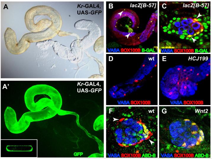
Figure 2. SOX100B-expressing cells give rise to testis PCs.
St. 17 gonads and adult testes immunostained as indicated in the figure. Anterior is to the left in each panel. (A,A’) Kr-GAL4, UAS-GFP adult testis examined by either light microscopy (A) or immunofluorescence (B). Inset: Slice of three-dimensional reconstruction of testis. Note that GFP expression is coincident with the yellow pigmentation of the testis and is only in the outer layer of cells. (B,C) The B-57 enhancer trap labels PCs in the adult testis (B) and st. 17 embryonic male gonad (C), and co-localizes with SOX100B (arrowheads). (D,E) Adult testes showing SOX100B-positive PCs in both wild-type (D) and AbdB[HCJ199] mutants (E). The AbdB[HCJ199] mutant testes do not have the normal, elongated appearance of wild-type testes, likely due to defects in interaction with the genital disc. (F,G) St. 17 embryonic male gonads immunostained to reveal msSGPs (co-expressing ABD-B and SOX100B) and PCs (SOX100B only, arrowheads). In addition, a few posterior SGPs also co-express ABD-B and SOX100B (DeFalco et al., 2003). Note that PCs are observed in wild-type (F) but not Wnt2 mutants (G).

