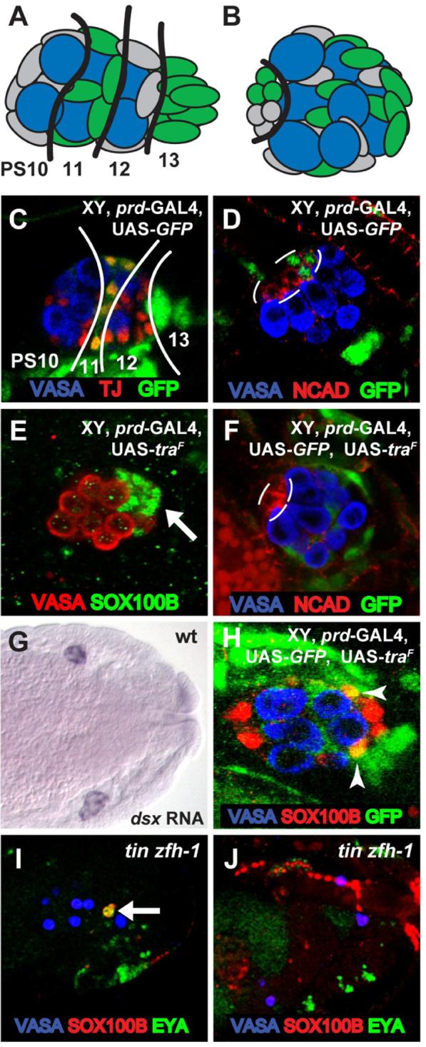
Figure 5. Non-cell autonomous control of sex determination in the Drosophila gonad.

(A,B) Diagram of prd-GAL4 expression (green) in the embryonic gonad at st. 15 (A) and st. 17 (B). Anterior is to the left in each panel. Approximate PS numbers are indicated in (A) based on the known pattern of prd gene expression. The black line in (B) outlines the hub. (C-F, H-J) Immunostainings of embryonic gonads as indicated. (C) GFP expression from st. 15 male embryo expressing prd-GAL4 and UAS-GFP. Traffic Jam (TJ) staining labels PS10-12 SGP nuclei. Lines separate GFP-positive and GFP-negative regions of the gonad. Note that GFP is expressed in PS 11 SGPs and strongly in the msSGPs from PS 13. GFP is also expressed in subsets of cells surrounding the gonad. (D) GFP expression from st. 17 male embryo expressing prd-GAL4 and UAS-GFP. Note that the hub (outline, labeled with DN-cadherin) is normally composed of GFP-positive (PS11) and GFP-negative (PS10) cells. (E) A similar embryo as in (C) but expressing TRA instead of GFP. Note that the msSGPs are still present in the XY gonad even though they express TRA. (F) A similar embryo as in (D) but now expressing TRA in addition to GFP. Note that GFP/ TRA expressing cells do not contribute to the hub (outline), in contrast to GFP-alone expressing cells in (D). (G) St. 15 wild type embryo labeled by in situ hybridization for dsx mRNA. (H) St. 17 embryo expressing prd-GAL4, UAS-GFP and UAS-traF, immunostained for SOX100B and GFP. Note that GFP/ TRA expressing cells (arrowheads) can still become PC precursors based on their expression of SOX100B and their position and morphology. (I,J) tin zfh-1 double-mutant embryos. Note that msSGPs (arrows, co-labeled with EYA and SOX100B) are present at st. 12 (I), but not at st. 15 (J).
