
Figure 4. Molecular marker analysis of migratory limb myoblasts and dorsal-ventral neural tube patterning.
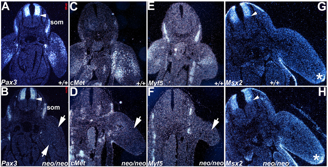
Radioactive in situ hybridization was used to assess Pax3 (A,B), cMet (C,D), Myf5 (E,F) and Msx2 (G,H) spatiotemporal expression patterns in E10 embryos. (A,B) In contrast to wildtype embryos (A), Pax3 mRNA is expressed at reduced levels within Pax3neoo/neo (neo/neo) dorsal NT (arrow head), adjacent dorsal root ganglia, somites (som) and is mostly undetectable within the hypomorphic limb buds apart from a few isolated myoblasts (B). The arrows indicate the two Pax3-expressing populations missing in hypomorphic limb buds. (C,D) cMet mRNA exhibits reduced expression in the Pax3neo/neo limb buds (arrow in D) when compared to wildtype (+/+) littermates (C). (E,F) Myf5 mRNA is also reduced in Pax3neo/neo limb buds (arrow in F) but is maintained in Pax3neo/neo somites. (G,H) Msx2 mRNA exhibits similar expression patterns in the dorsal NT, limb apical ectodermal ridge (*) and body wall within both wildtype (G) and hypomorphic embryos (H). Arrow heads points to mid-NT boundary.