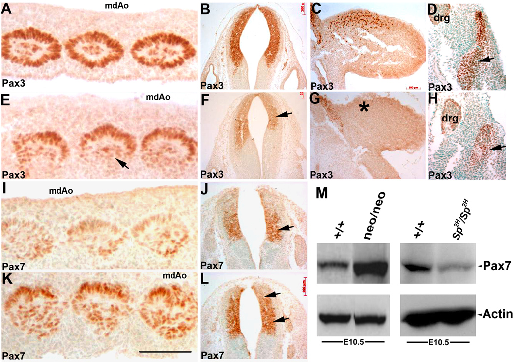
Figure 6. Pax3 protein levels are uniformly reduced whilst Pax7 protein levels are upregulated in Pax3neo/neo hypomorphs.

Immunodetection of Pax3 (A–H) and Pax7 (I–L) spatial expression and Western blot analysis (M) were used to assess relative Pax3 and Pax7 protein expression levels. In equivalent transverse sections through the midline dorsal aorta (mdAo) region, Pax3 protein is present in all the mesodermal cells of the concentric whorls of the E10.5 wildtytpe somites (A) but is expressed at reduced levels and is almost absent within the dorsolateral portion (arrow in E) of the Pax3neo/neo somites. Pax3 expression is also reduced in the E10.5 hypomorphic NT (F) and is absent within the E10.5 forelimb buds (G) when compared to wildtype embryos (B,C). In E11.5 Pax3neo/neo dermomyotome, the lateral region is absent and thus Pax3 expression is diminished (H) when compared to controls (D). In adjacent sections Pax7 protein is only weakly expressed in wildtype E10.5 somites (I) and restricted to the medial portion of the wildtype NT (J), but in contrast Pax7 expression is elevated in the hypomorphic somites (K) and expanded into the dorsal region of the NT in Pax3neo/neo embryos (L, indicated by arrows). (M) Western analysis of E10.5 whole embryos revealed Pax7 levels were elevated (~6 fold) in Pax3neo/neo embryos when compared to wildtype littermates, despite the actin loading control being expressed at similar levels. In contrast, Pax7 levels were reduced in Sp2H/Sp2H mutants when compared to wildtype littermates. Bar in K = 100 µm.
