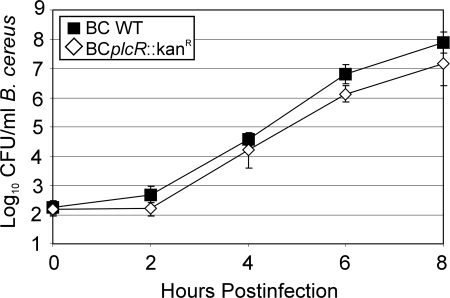
FIG. 1.
Growth of B. cereus in the presence of ARPE-19 cells. ARPE-19 cell monolayers were infected with 102 CFU/ml BCWT (▪) or BCplcR::Kanr (⋄) at the apical surface. Bacterial concentrations were quantified at 0, 2, 4, 6, and 8 h postinfection, and the two strains grew at similar rates (mean ± standard deviation; six per group).

