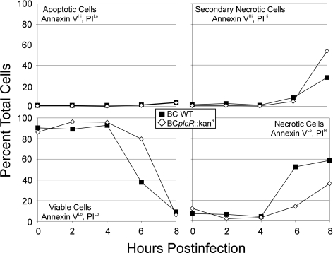
FIG. 3.
Necrotic death of RPE cells during infection with B. cereus. RPE cell monolayers were infected with 102 CFU/ml wild-type (▪) or plcR-deficient (⋄) B. cereus. At 0, 2, 4, 6, and 8 h postinfection, cells were harvested and treated with annexin V and PI to identify apoptotic and necrotic cells, respectively. This graphical representation shows that infected cell populations demonstrated a shift from viable to necrotic over time (represents six per group).

