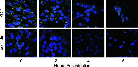
FIG. 6.
Changes in ZO-1 and occludin distribution in RPE cell monolayers in response to B. cereus infection. RPE cell monolayers were infected with 102 CFU/ml B. cereus and analyzed by immunocytochemistry for ZO-1 or occludin. Slides were examined by confocal microscopy at ×100 magnification. ZO-1 redistribution away from the cell periphery began at 6 h, and expression was lost by 8 h postinfection. Occludin disruption began as early as 2 h postinfection, and expression was lost by 6 h postinfection. The images are representative of six or more individual experiments.

