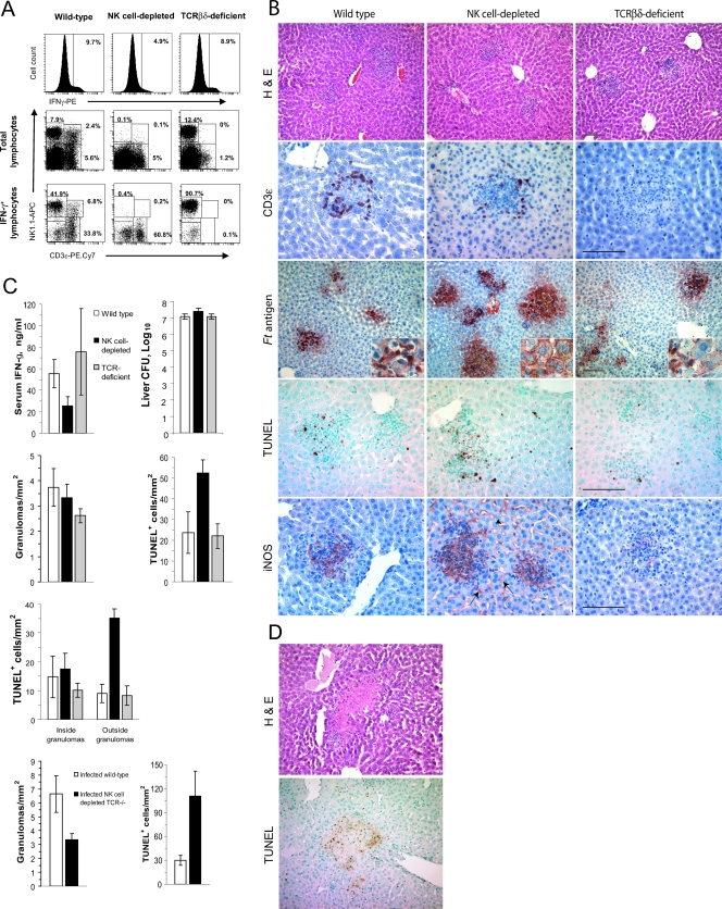
FIG. 6.
Depletion of NK cells alters the functions of hepatic granulomas in F. tularensis LVS-infected mice. Wild-type, NK cell-depleted, or T-cell-deficient mice (five per group) were each challenged i.n. with F. tularensis LVS, and samples of their livers were recovered 4 days later. (A) Hepatic mononuclear cells were analyzed by flow cytometry for the expression of intracellular IFN-γ (top row) and the presence of the major subsets of cells (middle row) or the three major subsets of lymphocytes or IFN-γ-producing cells (bottom row). PE, phycoerythrin; APC, allophycocyanin. (B) Granuloma formation and the distribution of CD3ɛ-expressing cells, Francisella antigens (Ft antigen), TUNEL-positive cells, and iNOS in the livers of infected mice. Note the presence of Francisella antigens within hepatocytes of NK cell-depleted mice and the expression of iNOS by Kupffer cells (arrow) and sinusoidal endothelial cells (arrowhead) of these animals. H&E, hematoxylin and eosin staining. (C) Serum IFN-γ concentrations, bacterial counts in the livers, and distribution of TUNEL+ cells in infected mice. Mice depleted of NK cells had significantly more bacteria in their livers, higher totals of TUNEL+ cells, and more TUNEL+ cells outside the granulomas than did wild-type mice (P < 0.05). Mice lacking NK, NKT, and T cells showed a significant decrease in the frequency of granulomas (P < 0.05) and a significant increase in the frequency of TUNEL+ cells in their livers (P < 0.05). (D) Dying cells within the livers of NK cell-depleted mice were found within areas of focal necrosis.

