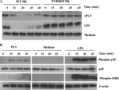
FIG. 7.
Activation of TLR4 downstream signals by rPLY. Peritoneal macrophages from C57BL/6 (WT) and TLR4 KO mice were stimulated with medium alone, rPLY, or LPS (1 μg/ml). (A) At the indicated times, cell lysates were collected and IκBα degradation was analyzed by Western blotting. (B) The cell lysates described in panel A were then subjected to Western blotting using antibodies specific to p38, phospho-p38, phospho-ERK, and β-actin. The results are representative of at least three independent experiments.

