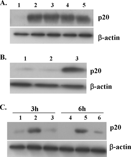
FIG. 9.
Activation of caspase-1 by rPLY and Streptococcus pneumoniae infection. Peritoneal macrophages were incubated with biotinylated YVAD-cmk (30 μM) for 1 h and subsequently stimulated with rPLY, cholesterol-treated rPLY, or LPS for 3 h, using different concentrations of rPLY or LPS. Activated caspase-1 was precipitated using streptavidin beads. Precipitates were subsequently analyzed for the presence of active caspase-1 (p20 subunit) by Western blotting using a caspase-1 antibody (A and B). (A) Lanes: 1, unstimulated; 2, 1 μg/ml rPLY; 3, 0.1 μg/ml rPLY; 4, 1 μg/ml cholesterol-treated rPLY; 5, 0.1 μg/ml cholesterol-treated rPLY. (B) Lanes: 1, unstimulated; 2, 1 μg/ml LPS; 3, LPS plus ATP (1 mM). (C) Similarly, peritoneal macrophages were incubated with biotinylated YVAD-cmk (30 μM) for 1 h and infected with wt S. pneumoniae and the Δply mutant at an MOI of 10, and cell lysates were collected at the indicated time points. Activated caspase-1 was detected as mentioned above. Lanes 1 and 4, uninfected cells; lanes 2 and 5, wt S. pneumoniae-infected cells; lanes 3 and 6, Δply mutant-infected cells. For all panels, results are representative of at least three separate experiments.

