FIG. 4.

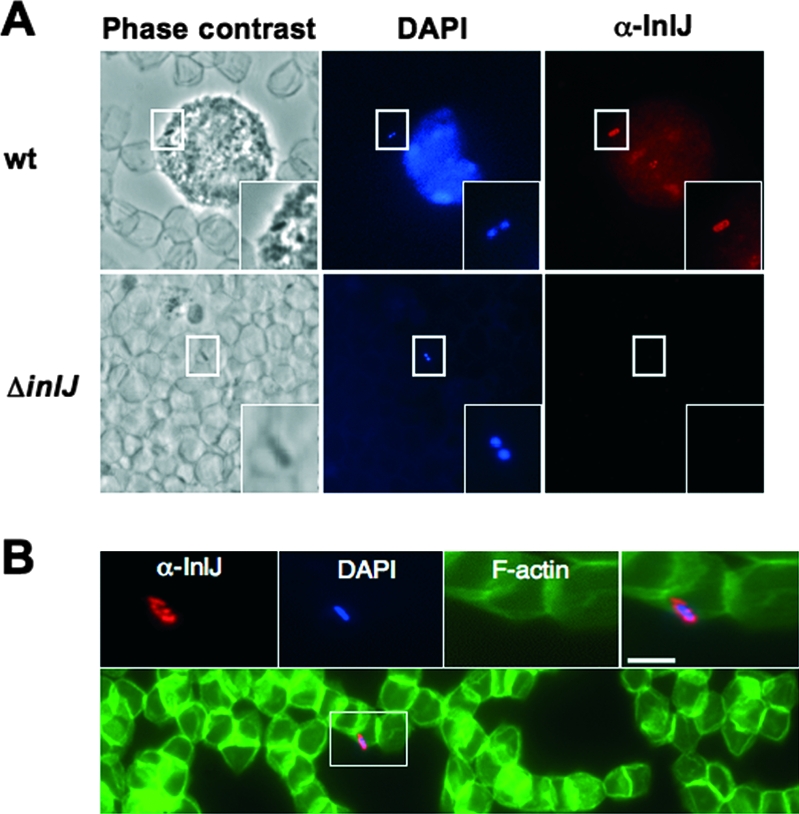
Bacteria present in the blood of infected mice produce InlJ. L. monocytogenes wild-type (wt) or ΔinlJ strain bacteria were inoculated intravenously into BALB/c mice. Following 72 h of infection, blood was recovered and fixed blood streams were labeled with InlJ antibody (in red) and DAPI, which detects white blood cells and bacterial DNA (in blue). (A) InlJ is present at the surface of a wild-type bacterium associated with a polynuclear cell (labeled with DAPI) and is absent at the surface of an inlJ mutant bacterium associated with erythrocytes (visible in phase-contrast image). (B) A wild-type L. monocytogenes bacterium in contact with erythrocytes expresses InlJ at its surface. Fluorescent phalloidin-488 labels actin in erythrocytes (in green). Scale bar, 2 μm. Rectangular regions indicate the positions of the magnified inset fields. α, anti-.
