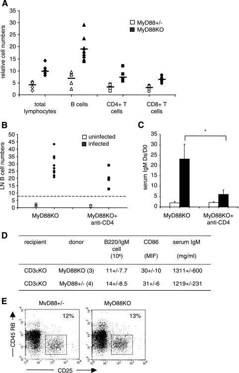
FIG. 6.
Although it is dependent on CD4+ T-cell function, IgM hypergammaglobulinemia observed in infected MyD88 KO mice is not linked to the absence of MyD88 in T cells. (A) Numbers of CD4+ and CD8+ cells in LN of infected MyD88 KO (black symbols) and MyD88+/− (white symbols) animals, determined by flow cytometry, relative to uninfected controls. Each point represents one infected animal compared to the noninfected control mean value. (B) MyD88 KO mice were injected intraperitoneally with an anti-CD4 monoclonal antibody (YTS177) to block CD4+ cells for at least 4 weeks (see Materials and Methods). Each point represents a mouse. Infected and treated MyD88 KO mice were compared with infected and nontreated MyD88 KO mice. Uninfected mice were used as controls. (C) Serum IgM levels in anti-CD4-treated and untreated MyD88 KO mice 4 weeks after infection with B. burgdorferi. Bars represent the mean ratios ± SD of the IgM concentrations between the day of sacrifice (Ds; 3 to 4 weeks after infection) and day 0 (D0) in anti-CD4-treated or untreated, uninfected (white) or infected (black) MyD88 KO animals. Differences between anti-CD4-treated and untreated infected MyD88 KO mice are significant (*, P = 0.009; Wilcoxon test). (D) Purified T cells (anti-CD11b, anti-CD45R, anti-DX5, and anti-Ter119 negative sorting; 20 × 106 cells) from MyD88+/− and MyD88 KO mice were transferred to CD3ɛ ΚΟ mice (three and four per group). After 24 h, the recipient mice were infected with B. burgdorferi and analyzed 3 weeks after infection. All mice displayed significant numbers of transferred CD4 T cells (6%) in LN. The basal level of IgM in noninfected CD3 KO animals was 342 ± 10 μg/ml. (E) Regulatory T cells in uninfected MyD88 KO and MyD88+/− mice. LN cells were gated on forward scatter and side scatter parameters. Numbers indicate percentages of CD25high CD45RBlow cells among CD4+ cells.

