FIG. 2.
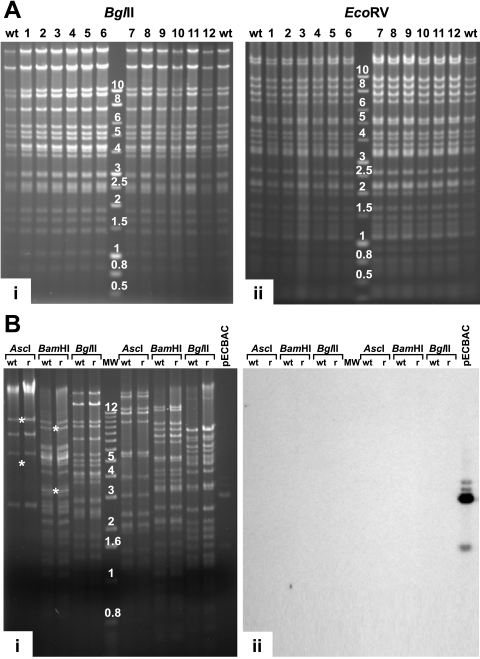
Restriction enzyme analysis of the rCCV genomes. (A) Parental wt CCV DNA (wt) and DNA of 12 independently regenerated and plaque-purified rCCV isolates (lanes 1 to 12) were digested with BglII (i) and EcoRV (ii) and separated by agarose gel electrophoresis. The molecular weight marker used was a 1-kb DNA ladder (Promega Corp.). (B) Restriction enzyme digestion and Southern blotting of rCCV-1 DNA. wt CCV (wt) and rCCV-1 DNA (r) were digested, separated by agarose gel electrophoresis (i), and hybridized with fragments of vector pECBAC1 (ii). *, AscI (5,597 and 10,224 bp) and BamHI (8,949 and 3,487 bp), DNA fragments which were derived from the termini of the linear virus demonstrating that rCCV-1 DNA isolated from virions contained both direct terminal repeats. pECBAC1, digested using PstI (1,541-, 2,949-, and 3,029-bp fragments), was used as a positive control. MW, molecular weight marker with band size in kb (1-kb DNA ladder; Invitrogen Inc.).

