FIG. 8.
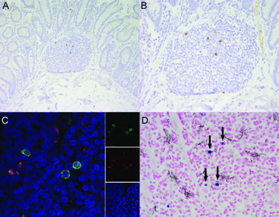
Tissue distribution of SIVmac239ΔV1V2 during acute viremia. (A and B) Localization by immunohistochemistry of SIVmac239ΔV1V2 in gut-associated lymphoid tissue revealed viral infection primarily of organized lymphoid nodules and absence from the lamina propria. (C) Colocalization of SIV and CD68 in the mesenteric lymph node of SIVmac239ΔV1V2 inoculated rhesus macaque revealed that 93.5% of SIV-infected cells are also CD68 positive. Images for individual panels (red, CD68 [KP-1]; green, SIV nef; and blue, ToPro-3 nuclear marker) are shown on the right and merged to form a composite image on the left. Yellow-to-orange areas indicate colocalization of SIV and CD68. (D) Colocalization failed to demonstrate that DC-SIGN-expressing dendritic cells were targeted. SIV-infected cells demonstrated by in situ hybridization are in blue (arrows); DC-SIGN-positive dendritic cells demonstrated by immunohistochemistry are in black.

