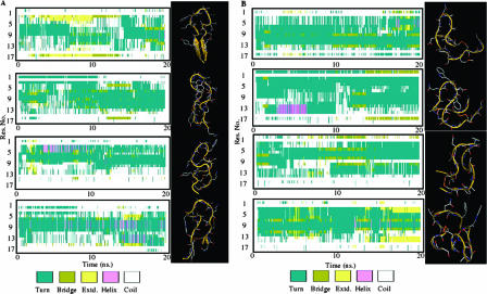
FIG. 5.
Secondary structure and models of Ala95-96 and cyclic WT peptides. (A) Structure and model of the WT peptide. The left side shows the time evolution of the secondary structure of the WT peptide in each of the four MD simulations. For each frame of the trajectory, the algorithm STRIDE assigned the secondary structure of each residue of the peptide. Different secondary-structure motifs are identified by the color code depicted in the legend. The right side shows a representation of the most populated structure of the peptide extracted by the cluster analysis of each trajectory. (B) Structure and model of the Ala95-96 peptide. The left side shows the time evolution of the secondary structure of the peptide in each of the four MD simulations. For each frame of the trajectory, the algorithm STRIDE assigned the secondary structure of each residue of the peptide. Different secondary-structure motifs are identified by the color code depicted in the legend. The right side shows a representation of the most populated structure of the peptide extracted by the cluster analysis of each trajectory.

