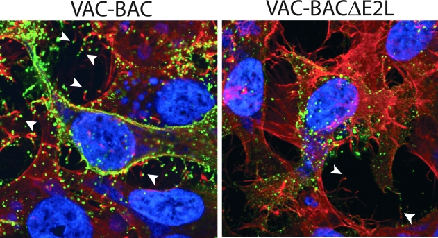
FIG. 11.
Visualization of EVs with actin tails. Cells infected with VAC-BAC or VAC-BACΔE2L were fixed with 3% paraformaldehyde and quenched with 2% glycine. EVs were labeled with MAb to the B5 protein and Cy5-conjugated goat anti-rat immunoglobulin G secondary antibody. Following this, cells were permeabilized by the addition of 0.1% Triton X-100 and stained with 4′,6-diamidino-2-phenylindole to visualize DNA and with Alexa Fluor 568 phalloidin to visualize filamentous actin. Arrowheads indicate representative EVs at the tips of actin tails.

