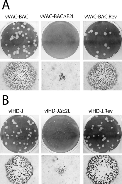
FIG. 4.
Plaque sizes of ΔE2L viruses. (A) Comparison of ΔE2L viruses in the VACV WR background. vVAC-BAC, vVAC-BACΔE2L, and vVAC-BAC.Rev were plated on BS-C-1 cells under semisolid medium. After 4 days, plates were stained with crystal violet to visualize a field of plaques (top row) or fixed with acetone-methanol (1:1) and stained with anti-VACV antibody 8191 followed by anti-rabbit immunoglobulin G conjugated to peroxidase to visualize individual plaques by microscopy (bottom row). (B) Comparison of ΔE2L viruses in the VACV IHD-J background. vIHD-J, vIHD-JΔE2L, and vIHD-J.Rev were analyzed as described for panel A.

