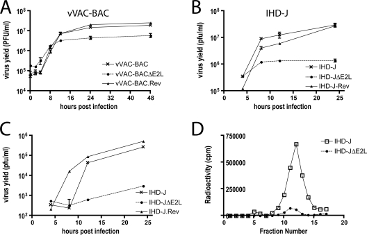
FIG. 6.
Intracellular and extracellular virus production. (A and B) Infectious-MV formation. BS-C-1 cells were infected with 5 PFU per cell of vVAC-BAC, vVAC-BACΔE2L, or vVAC-BAC.Rev or with the same amount of vIHD-J, vIHD-JΔE2L, or vIHD-J.rev. At the indicated times after infection, cells from individual triplicate wells were harvested and disrupted, and the viral yields were measured by plaque assays on BS-C-1 cells. Standard error bars are shown. (C) Infectious-EV formation. The media from triplicate wells were individually harvested at the indicated times and clarified by low-speed centrifugation. Contaminating MVs were neutralized with MAb 7D11 to the L1 protein (43) prior to plaque assay, as for panels A and B. (D) Analysis of metabolically labeled EV by CsCl gradient sedimentation. RK13 cells were infected with vIHD-J or vIHD-JΔE2L and labeled with [35S]cysteine and [35S]methionine. The medium was collected at 18 h and clarified by low-speed centrifugation. The supernatant material was purified by sedimentation through a 36% sucrose cushion followed by CsCl gradient centrifugation. Fractions were collected from the bottom of the tube, and radioactivity was determined by scintillation counting.

