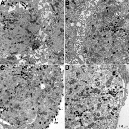
FIG. 8.
Electron-microscopic images of cells infected with viruses containing or lacking the E2L ORF. Thin sections of cells infected with vVAC-BAC (A), vVAC-BACΔE2L (B), vIHD-J (C), or vIHD-JΔE2L (D) are shown. Representative immature virions (IV), MVs, and EVs are indicated with arrows. The junction between two cells appears in panels A and B.

