
FIG. 6.
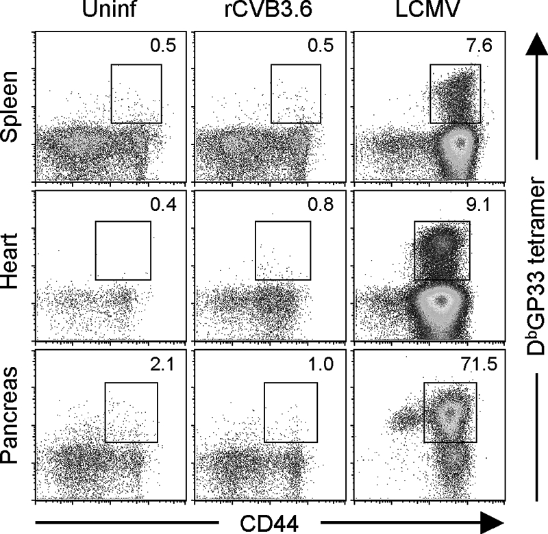
CVB3-specific CD8+ T cells cannot be readily identified in lymphoid or nonlymphoid tissues by using MHC class I peptide tetramers. Mice were infected with rCVB3.6 or with LCMV, and 8 days later, the mice were perfused and sacrificed. Splenocytes and mononuclear cells isolated from the heart and pancreas were stained with CD8 and CD44 antibodies and MHC class I H-2Db/GP33-41 tetramers and analyzed by flow cytometry. Cells isolated from the hearts and pancreases of three mice per group were pooled for analysis. The dot plots shown are gated on CD8+ T cells, and the numbers indicate the percentages of CD8+ T cells that were tetramer positive. Uninf, uninfected.