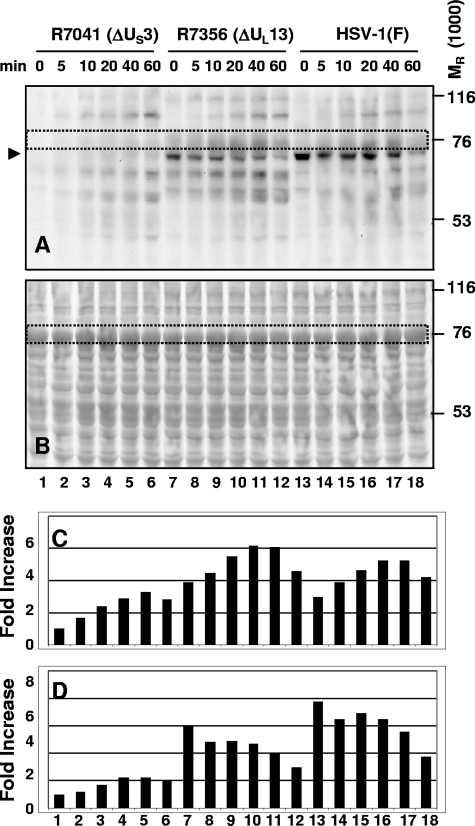
FIG. 3.
GST-cdc25C-M chimeric protein is phosphorylated by infected-cell lysate in the presence of US3. Lysates of HEp-2 cells harvested 12 h after infection with ΔUS3, ΔUL13, or wild-type HSV-1(F) virus were incubated with GST-cdc25C-M chimeric protein for a total of 60 min in kinase reaction buffer containing [γ-32P]ATP, spending the indicated time of 0, 5, 10, 20, 40, or 60 min at 30°C after being incubated on ice for 60, 55, 50, 40, 20, or 0 min, respectively. The mixtures were electrophoretically separated on a 10% denaturing gel, transferred to a membrane, and visualized by autoradiography (A). The blot was then stained with Ponceau S to detect total protein levels (B). The GST-cdc25C-M band is outlined by a dotted rectangle and was quantified as shown in panel C. The 70-kDa band consistent with ICP22 is marked by an arrowhead and was quantified as shown in panel D. Quantification of 32P phosphorylation of the substrate was done using a Molecular Dynamics PhosphorImager. The quantification of the amounts of radioactivity in each band was normalized with respect to the amount of radioactivity present in the R7041-infected lysate at 0 min (lane 1).

