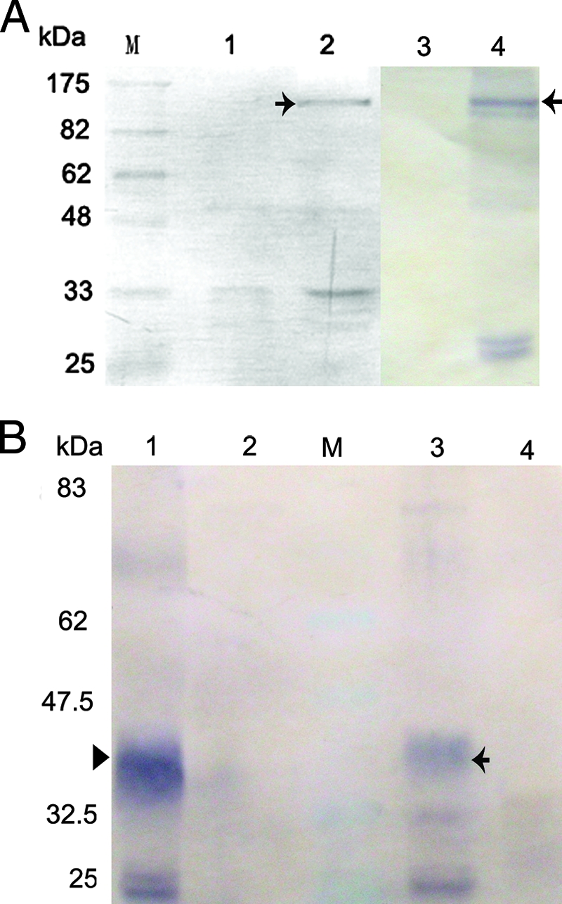
FIG. 6.

Immunoprecipitation analysis of the interactions between VP48R and FLK-1. Lane M is molecular size markers. (A) The expression vector PC-48V5 together with the expression vector PN-flk or the empty pEGFP-N3 vector were used to cotransfect FHM cells. Cytoplasmic extracts from these cells were immunoprecipitated with an anti-V5 monoclonal antibody and detected by Western blotting using the anti-GFP antibody. (Lane 1) As a control, no band corresponding to the GFP (28K) was detected in the immunoprecipitation analysis by using cells cotransfected with PC-48V5 and pEGFP-N3 vectors. (Lane 2) FLK-GFP protein (black arrow) was coprecipitated by VP48R. (Lane 3) Lysates of FHM cells showed no positive signals to anti-GFP antibody. (Lane 4) Western blot analysis showed the band corresponding to the FLK-GFP protein in the lysates of FHM cells cotransfected with PN-flk and PC-48V5. (B) The expression vector PC-flkV5/PN-48 or PC-flkV5/pEGFP-N3 was used to cotransfect FHM cells. Cytoplasmic extracts from these cells were immunoprecipitated with an anti-V5 monoclonal antibody and detected by Western blotting using the anti-GFP antibody. (Lane 1) Lysates of FHM cells showed no positive signals to anti-GFP antibody. (Lane 2) Western blot analysis showed the band corresponding to the FLK-GFP protein in the lysates of FHM cells cotransfected with PN-48 and PC-flkV5. (Lane 3) The band corresponding to the 48-GFP protein (40.5K) (black arrowhead) was detected in the immunoprecipitation analysis by using FHM cells cotransfected with PC-flkV5 and PN-48 vectors. (Lane 4) As the control, no band corresponding to the GFP (28K) was detected in the immunoprecipitation analysis by using FHM cells cotransfected with PC-flkV5/pEGFP-N3.
