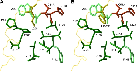
FIG. 1.
The structure of the FMDV S2 pocket of Lbpro. The side chains of the wild-type P2 residue L200 (A) and the modeled L200F mutation (B) after energy minimization are shown. Amino acid side chains comprising the pocket are dark green. Light-green residues contribute to the formation of the S2 pocket through main-chain interactions. Active-site residues are shown in red (C51 was replaced by alanine in the crystal structure (PDB code 1QOL) (10). The backbone of the C-terminal extension of the adjacent molecule is in yellow.

