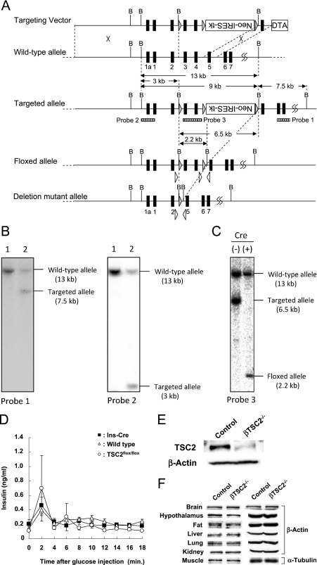
FIG. 1.
Generation of β-cell-specific TSC2 knockout mice. (A) Structure of targeting vector and Tsc2 alleles. Exons are denoted with filled boxes, some with numbers. Expression cassettes for the diphtheria toxin A chain (DTA) and Neo/HSV-TK (Neo-IRES-TK) are shown as open boxes. Shaded triangles indicate loxP sequences. Dotted lines indicate sites of insertion (targeting vector into wild-type allele) and excision (targeted allele reduced to floxed allele and reduced again to deleted mutant allele). Open triangles indicate primers for genotyping. Positions of probes used for Southern blot analysis are shown as striped boxes below the targeted allele. B; BamHI restriction site. (B) Southern blot analysis of G418-resistant ES clones. BamHI-digested DNAs from two ES clones were analyzed with two probes. Lane 1, nonhomologous recombinant control clone; lane 2, homologous recombinant clone. (C) Southern blot analysis of the floxed Tsc2 allele. BamHI-digested DNA was analyzed. Cre (−), parental homologous recombinant ES clone; Cre (+), FIAU-resistant ES clone with floxed allele. (D) Time course of changes in plasma insulin concentration induced by intraperitoneal administration of glucose (3 mg per gram of body weight) in wild-type (open triangles; n = 3), Ins-Cre (filled squares; n = 3), and TSC2flox/flox (open circles; n = 4) mice at 8 weeks of age. Data are means ± SEM. (E, F) Immunoblot analysis of TSC2 and either β-actin or α-tubulin (loading controls) in pancreatic islets (E) or in the indicated tissues (F) of 9-week-old control (TSC2flox/flox) and βTSC2−/− mice.

