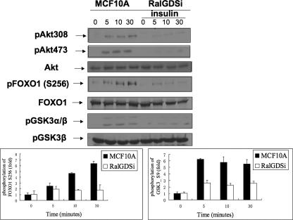
FIG. 2.
Signaling downstream from Akt is suppressed by RalGDS knockdown in MCF10A cells. MCF10A cells stably expressing empty vector or RalGDS shRNA were serum starved for 6 h and then stimulated with insulin (1 μM) for the indicated times, and cell lysates were assayed for pAkt308, pAkt473, total Akt, pFOXO1 (Ser256), total FOXO1, pGSK3α/β (Ser21/9), and total GSK3β. pFOXO1 and pGSK3β band intensities were quantified with NIH ImageJ 1.34s, and the data represent the averages of data from three independent experiments.

