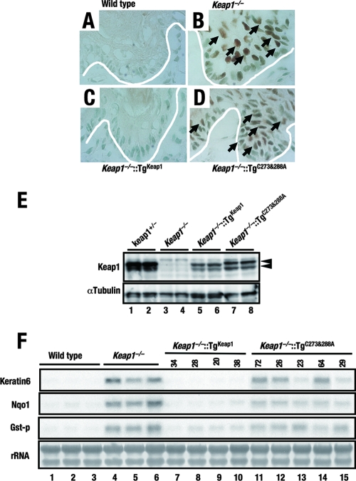
FIG. 6.
Constitutive nuclear accumulation of Nrf2 caused by the C273A-C288A mutation of Keap1. (A to D) Immunohistochemical staining of forestomach epithelia of wild-type (A), Keap1−/− (B), Keap1−/−::TgKeap1 (C), and Keap1−/−::TgC273A-C288A (D) mice with anti-Nrf2 antibody. White lines indicate the positions of the basement membranes. Brown staining can be seen in the nuclei of cells from Keap1−/− and Keap1−/−::TgC273A-C288A mice (arrows). (E, top panel) Immunoblot analysis showing the expression levels of Keap1 and the Keap1(C273A-C288A) mutant form by using anti-Keap1 antibody. Whole-cell lysates of forestomachs derived from Keap1−/−::TgKeap1 (line 38) and Keap1−/−::TgC273A-C288A (line 64) mice were analyzed. Arrowheads indicate Keap1. (E, bottom panel) α-Tubulin was used as an internal control. (F) RNA blot analysis of total RNAs purified from forestomachs. The abundances of keratin 6, Nqo1, and GST-P mRNAs were examined, and these mRNAs were shown to be highly expressed in Keap1−/− and Keap1−/−::TgC273A-C288A mice. rRNA was utilized as an internal control.

