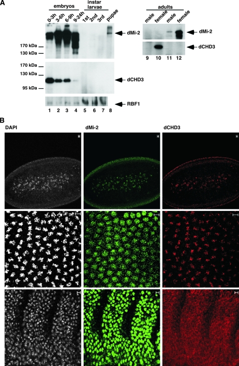
FIG. 6.
dCHD3 expression in embryos. (A) Extracts derived from different developmental stages were immunoprecipitated and subjected to Western blot analysis as indicated using the following antibodies: for dMi-2, anti-dMi-2-N; for dCHD3, 5; and for RBF1, DX3. Antibodies used for immunoprecipitation were, for lanes 1 to 8, anti-dMi-2 (4D8, upper panel), anti-dCHD3 (7A11, middle panel), and anti-RBF1 (DX3, bottom panel); for lanes 9 and 10, anti-dCHD3 (7A11); and for lanes 11 and 12, anti-dMi-2 (4D8). Antibodies used to probe the Western blots are indicated on the right. (B) Immunofluorescence. Early preblastoderm (upper panels), late preblastoderm (middle panels), and postgastrulation (lower panels) embryos were stained with DAPI (white; left panels), dMi-2 (green; middle panels), and dCHD3 antibody (red; right panels). Scale bars, 5 μm.

