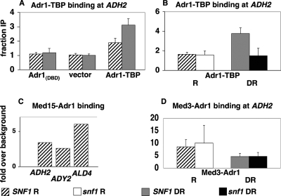
FIG. 1.
Binding of Adr1 DBD fusions to Adr1-dependent promoters. ChIP was performed and the results were quantitated as described in Materials and Methods with strains with the indicated Adr1 DBD fusions or controls. (A) ChIP for the Adr1 DBD only (plasmid pAdr1Δ172 [17]), negative control vector (pKD8 [17]), or Adr1-TBP (Materials and Methods), all in ECY1 and grown under repressing conditions (hatched bars) or after 4 h of derepression (gray bars). Data are expressed as the ratios described in Materials and Methods. (B) ChIP of Adr1-TBP in a SNF1 strain (TYY497), repressed (R) (hatched bar) or 4-h derepressed (DR) (gray bar), and a snf1Δ strain (TYY498), repressed (white bar) or 4-h derepressed (black bar). Data are expressed as described for panel A. (C) ChIP of Med15-Adr1 under repressing conditions using strain ECY5. Data are expressed as increases over background from an untagged, repressed adr1Δ strain (ECY1). (D) ChIP of Med3-Adr1 in repressed SNF1 (ECY10) (hatched bar) and snf1Δ (CTYTY75) (white bar) strains and in the same strains after 4 h of derepression (gray and black bars, respectively). Data are expressed as described for panel C. Error bars in panels A, B, and D indicate standard errors of the means for two or three biological replicates.

