Figure 7.

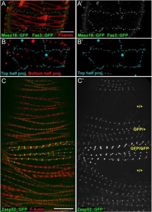
Epithelial sheath cells are mononucleate. (A-A’) Confocal projection of top half of an ovariole expressing both Fas3::GFP (green dots) and Meso18::GFP (green nuclei) in epithelial sheath cells stained with antibodies to FLN12−20 (red). (A’) A single cell is clearly outlined by Fas3::GFP dots and contains only one nucleus, as seen with Meso18::GFP. (B-B’) Rendering of optical sections taken through the entire ovariole shown in A. Only green signal from Fas3::GFP and Meso18::GFP was rendered. The top half of the confocal stack is colored cyan, the bottom half red. B’ shows the top half only, and represents a rendering of the raw data shown in A’. See Supplemental movies for rotation of the raw data and rendered stacks. (C-C’) Confocal image of an ovariole with Flp/FRT-induced mitotic clones. F-Actin is in red and Zasp52::GFP is in green. C’ shows Zasp52::GFP only, with the inferred copy number of GFP genes indicated in yellow. Scale bars: 20 μ.
