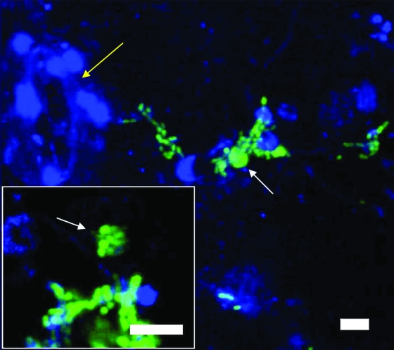
FIG. 8.
Confocal micrographs of vesicles (white arrows) containing cells of GFP-labeled S. enterica MB156. The vesicles were expelled by Tetrahymena sp. during grazing on the leaves of cilantro plants inoculated with the enteric pathogen and 24 h later with the protist. Note the presence of distinct green fluorescent bacterial cells in the vesicle shown in the insert. The autofluorescence of the plant tissue was assigned the pseudocolor blue. The yellow arrow indicates the presence of a stomate. Scale bars, 10 μm.

