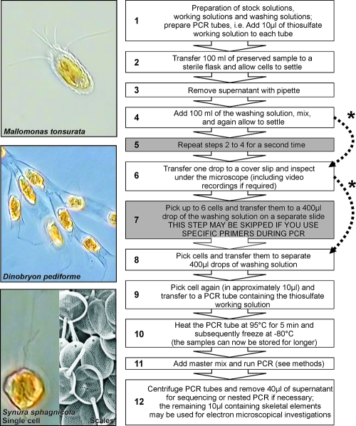
FIG. 2.
Proposed protocol. The essential steps of the protocol are depicted with a white background, those with a gray background may be skipped depending on the level of contamination/cell concentrations in the original sample and the specificity of the primers used during PCR (indicated by the dotted arrows marked with an asterisk). For an explanation, see the text. Examples of the morphological analysis are shown on the left for three species. For Synura sphagnicola, a scanning electron microscopy image of the scales, which can be extracted from the PCR tubes after running the PCR, is also shown.

