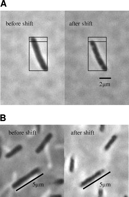
FIG. 6.
Cells of E. coli M2701 before and after an osmotic upshift. Cells of E. coli M2701 in K20 minimal medium that were attached to the microscope slide without fixation were photographed before and 10 s after an osmotic upshift by the addition of 0.8 M NaCl (A) or 0.8 M KCl (B) to the side of the coverslip. Pictures were taken at 1,000-fold magnification under a phase-contrast microscope. For easier size estimation, the subdivided squares of identical size or metering bars were introduced in the figure.

