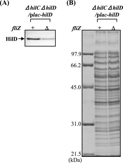
FIG. 7.
Cellular levels of HilD protein expressed by a PA1lacO-1 promoter-hilD fusion in wild type and FliZ-depleted cells. Plasmid pTKY651 (PA1lacO-1 promoter-hilD fusion) was introduced into bacterial strains CS2802 (fliZ+ ΔhilC ΔhilD) and CS3329 (ΔfliZ ΔhilC ΔhilD). Cells of the resultant strains were grown to an optical density at 600 nm of 1.0, collected, lysed, and run on SDS-10% polyacrylamide gels. The separated proteins were transferred to a membrane and then immunostained with anti-HilD antiserum (A). Coomassie brilliant blue-stained gel electrophoretic patterns of the same samples used for immunoblotting are also shown (B). The leftmost lane in panel B contains molecular mass standards.

