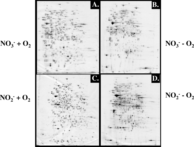
FIG. 1.
2-D gels of aerobically (A and C) and anaerobically (B and D) expressed P. aeruginosa proteins. Cell extracts (30 μg) derived from the same growth phase (see Fig. S1 in the supplemental material) grown with NO3− (A and B)- or NO2− (C and D)-containing medium were separated by 2-D gel electrophoresis and stained with silver nitrate. The numbered spots correspond to proteins identified by MALDI-TOF analysis (Table 1).

