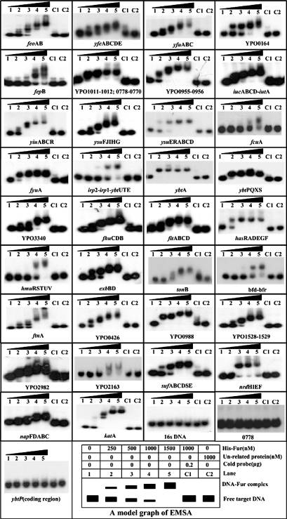
FIG. 2.
EMSAs. The PCR-generated, [γ-32P]-labeled target DNA probe or the corresponding cold probe (i.e., unlabeled target DNA) was incubated with the purified His-Fur protein or an unrelated protein (purified goat anti-rabbit anti-F1 antigen antibody). The mixtures were directly subjected to the 4% (wt/vol) PAGE. The band of free promoter DNA disappears with increasing amount of Fur protein, and a retarded DNA band with decreased mobility turns up, which presumably represents the Fur-DNA complex. A model graph of EMSA is shown as well.

