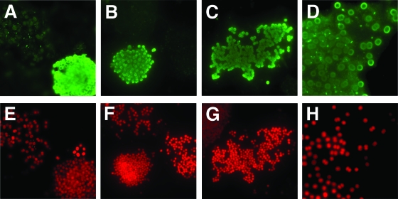
FIG. 7.
Immunofluorescence micrographs of Microcystis field colonies from samples obtained from Lake Dollgow (Germany) in 2007 produced by using the antibody against MrpC and a FITC-coupled secondary antibody. Images of cells were acquired either with a green fluorescence emission filter (A to D) in the blue channel of the microscope or with a red fluorescence emission filter (E to H) in the green channel of the microscope. Panels D and H show enlarged sections of an individual colony.

