
Figure 5. A PEG molecule is present in both monomers of the T57S-ethanol structure.
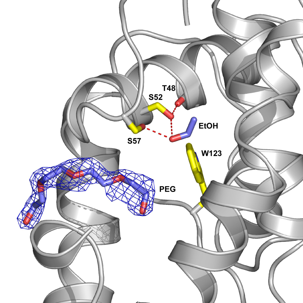
A region of a PEG molecule (blue) is observed to enter the site but does not interfere with alcohol binding. Yellow residues highlight the ethanol binding site and the ethanol is shown in blue. The protein was crystallized using 25–29% PEG4000, which contains ~ 90 repeating units, and the remainder of the PEG molecule, which is presumably outside of the binding site is not defined.