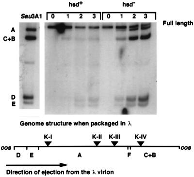
Figure 2.
Autoradiogram depicting the time course of methylation of T7 DNA ejected from a λ virion during the infection of the hsd+ strain IJ891(pTP166) and of the Δhsd strain IJ1133(pTP166). Time (in minutes) after infection is shown above the lanes. T71.7∷λcosΔ10-NB1[λ] was added to cells at a multiplicity of 0.4. The lane marked Sau3AI illustrates the pattern of cleavage when each GATC site in the phage genome is cut (GATC sites are present in the λ cos DNA; any circular form of intracellular T7 DNA caused by annealing and ligation of the single-stranded ends of T7[λ] DNA cannot be detected in this experiment). DNA in other lanes was cleaved by DpnI; fragments are indicated by letters to the left of the panel, and their location on the phage genome is indicated on the map below; Dam methylation sites (vertical bars) and EcoKI recognition sites (triangles) are also shown. The 103-bp G fragment is not indicated.

