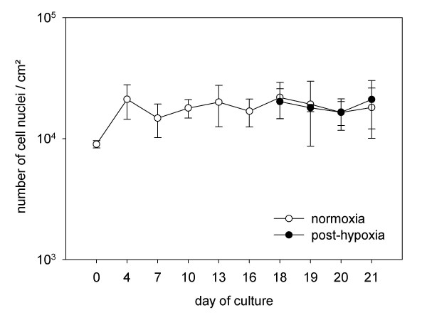
Figure 2.
Progression of the number of nuclei after differentiation period and effect of hypoxia on cell count. Nuclei were stained with DAPI and counted in fluorescence micrographs. Data were calculated from three independent experiments including multiple wells, each based on 25 micrographs. The white circles represent cultures that were cultivated under continuously normoxic conditions. The black circles illustrate the progression of the number of nuclei in post-hypoxic cultures. Under both conditions number of nuclei remained stable.

