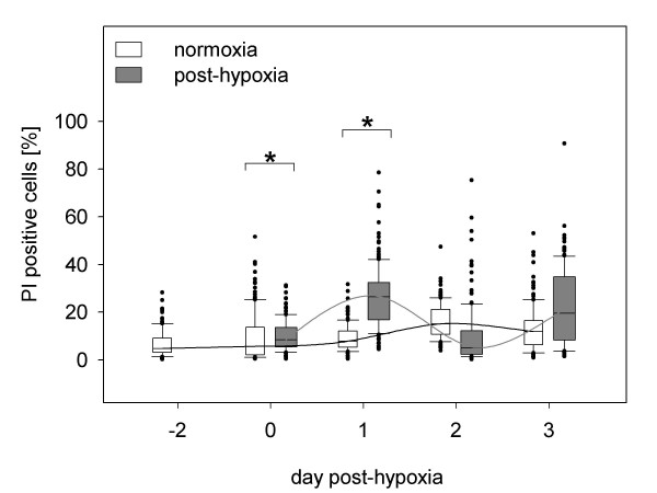
Figure 5.
The progression of PI positive cells in post-hypoxic and normoxic SH-SY5Y cultures. Results are derived from three independent experiments, in which 75 pictures were taken for each analysis. Significant increases in the rate of PI positive cells were observed only at Day 0 (10% ± 6%) and Day 1 (27% ± 13%) compared to normoxic cultures (Day 0: 9% ± 10%); Day 1: 9% ± 6%). Note PI stains necrotic and late apoptotic cells. No significant difference of necrosis rate was detected of post-hypoxic cultures as well as in control cultures during the observation period.

