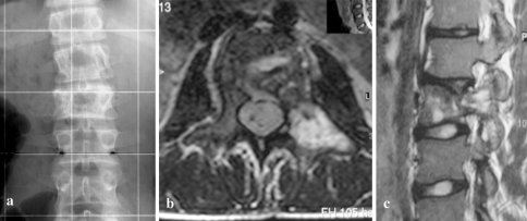
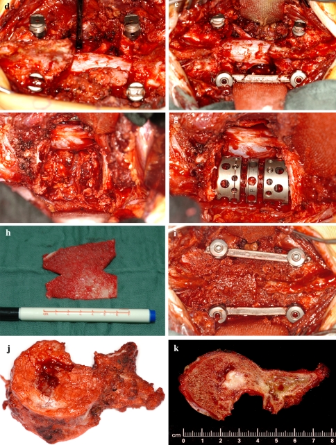
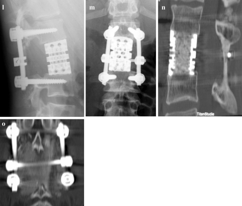
Fig. 2.
a–c 16-year-old male with Ewing’s sarcoma of L2 (patient number 6) and involvement of the left pedicle and transverse process. d Intraoperative view after resection of the healthy posterior structures to the base of the right pedicle with release of the right L2 nerve root. e and f Posterior and anterior view after en bloc spondylectomy. g Anterior column reconstruction with expandable titanium cage. h and i The cortical graft is trimmed and placed pressfit inbetween the spinous processes and covered with bone chips. j and k Macrospecimen and macrosection of L2 demonstrating clear margins. l and m Postoperative X-rays. n and o CT scans 3 years postop. showing bony fusion of both the cage and the posterior cortical graft


