
Figure 4.
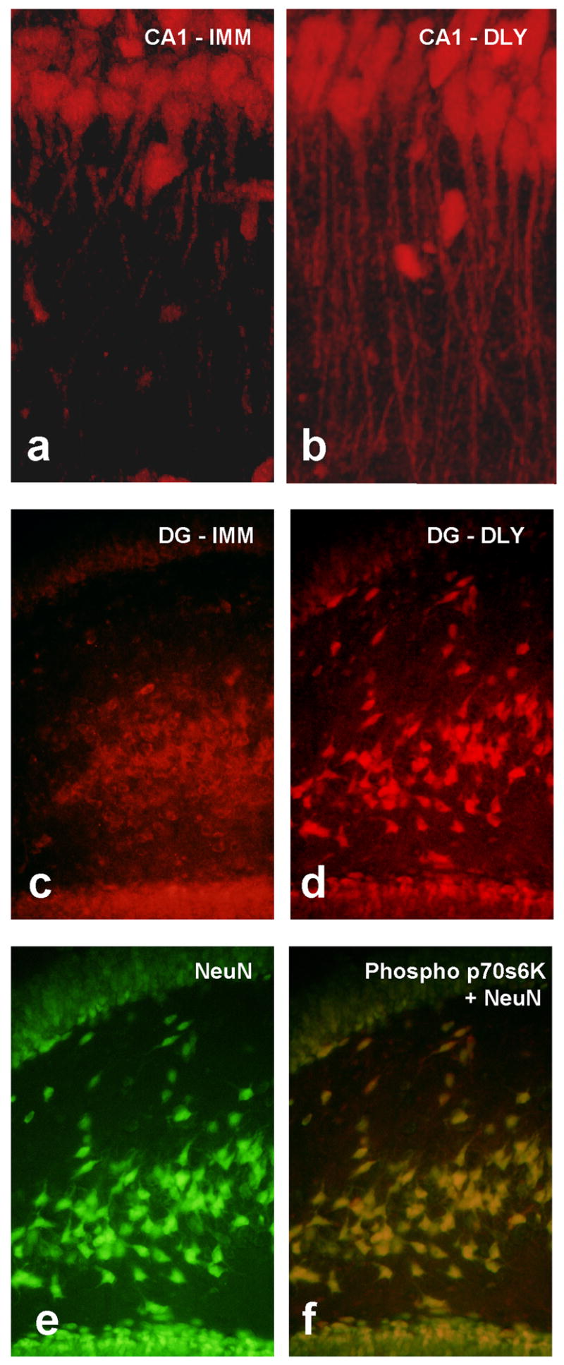
Immunofluorescence images of phosphorylated p70s6k in hippocampal tissue from rats given immediate (IMM) or delayed (DLY) shock after placement in a novel chamber. Confocal images show distinct patterns of staining in apical dendrites of CA1 pyramidal neurons in animals that learned (b) compared to those that did not (a) supporting the potential importance of mTOR-dependent translation in dendrites. Dentate gyrus showed a similar pattern in that more intense and more punctate staining was found in the DLY group (d). Double labeling with an antibody to the neuron-specific DNA binding protein NeuN (e) indicated that the p70s6k signal is seen primary in neurons. Quantitative protein assays on hippocampal tissue from these groups confirm these observations (see Gafford et al., 2007).