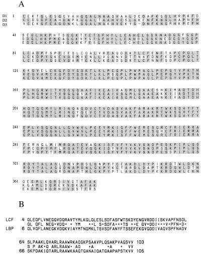
Figure 4.
(A) Peptide sequence alignment of the three repeated domains, with the consensus residues shaded. The first residue of each repeated domain is numbered 1. (B) Peptide sequence alignment of the N-terminal regions of G. polyedra LCF and LBP. The identical residues are shown in the middle line (between LCF and LBP sequences). The symbol + indicates that the residues at that position are different but functionally conserved, based on the blast pam 240 program (23).

