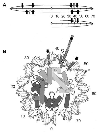
Figure 6.
Summary of cross-links. (A) Linear representation of cross-linking by one H2A N-terminal tail domain within 5S (Upper) or random sequence (Lower) nucleosome cores. Sites of cross-linking from cross-linking probe positioned at the second (H2AG2C) or 12th amino acid residue (H2AA12C) are indicated by the filled or open arrows, respectively. Distance in base pairs from the center (dyad) of the nucleosome is indicated. (B) Positions of cross-links by one H2A N-terminal tail domain within one superhelical turn of nucleosomal DNA. Top view of the nucleosome showing the locations of cross-links as in A. Only protein and DNA in the top half of the nucleosome are shown. Core histone α-helices (2) are represented by columns and other secondary structures as thin tubes as in Felsenfeld (27). The mobile histone tail domains are indicated by the dashed lines, and residues within the N-terminal tail of H2A are indicated by 3.5-Å spheres. The positions of the 2nd and 12 residues are indicated by the closed and open spheres, respectively. Histones H3, H4, H2B, and H2A are shaded dark to light gray, respectively. Note that a small portion of H3 from the bottom half of the octamer is shown. Numbers indicate distance from nucleosomal dyad as in A.

